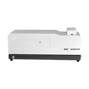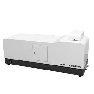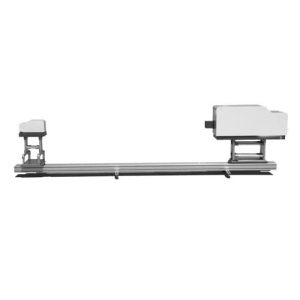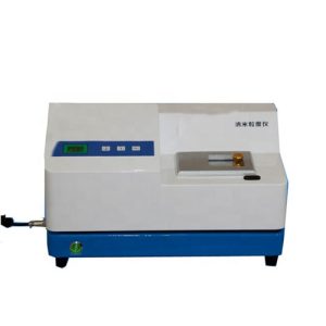

LR-T190 Microscope Particle Image Analyser
- Description
- Inquiry
Description
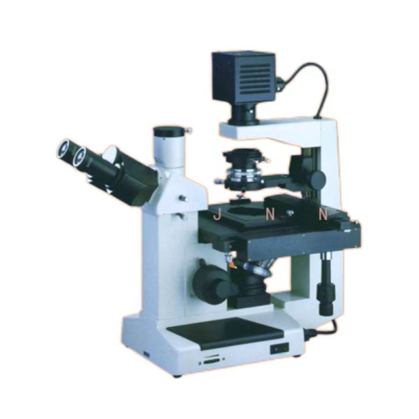
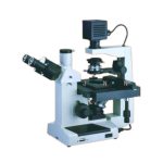
Microscope Particle Image Analyser
I. Product Introduction
The LR-T190 particle imager is a new generation of particle image analysis equipment developed by our company. The LR-T190 adopts the latest industrial-grade microscopic image acquisition system to make the sample observation clearer. The analysis software system can analyze and calculate the “monomer data”, “morphological parameters”, “overall distribution” and other parameters of the sample, and can easily obtain rich data such as the curve of the overall distribution of the sample and the description of the particle morphology. It is a professional instrument and equipment for the observation and analysis of all kinds of particle samples. The supporting professional particle analysis software has been upgraded and improved more humanized according to the user’s opinions over the years, and the computer independently analyzes the sample’s equivalent particle size, X, Y tangent and other “monomer properties”, as well as “morphological parameters” including aspect ratio and spherical degree. Through further calculation, the overall distribution of the sample (including the overall distribution curve and other rich data) is obtained. The software also increases the calculation mode of multiple image stitching, which increases the number of particles involved in the analysis, thus effectively ensuring the representaticity of the data. Finally, the sample data can be output in the form of a report (including image samples, charts, data lists, etc.), which is very convenient for testers to manage and report the test results.
Microscope Particle Image Analyser
Product advantages
As a professional particle image analysis instrument, LR-T190 particle image instrument is designed and developed for particles or particle related industries. Its outstanding advantages are mainly reflected in the following aspects:
※1, strong professionalism: Compared with various types of image analysis instruments produced by other manufacturers, our particle testing research experience and image theory are perfectly combined, which is the professional guarantee of “LR-T190 particle image instrument”. This product can not only scientifically describe the particle monomer of the sample, but also visualize the distribution of particles using data, charts and other ways. Can independently or assist laser particle size instrument and other equipment to better carry out particle testing work.
※2. The data is intuitive, easy to understand, and the analysis results are clear at a glance: common particle testing instruments include laser particle size meters, sedimentation particle size meters, etc., but the intuitive advantage of observing the sample morphology while conducting particle detection is not available in all other particle testing equipment, which can enable users to more comprehensive understanding of the particle morphology, state, change process and other information. Moreover, the comparison with the intuitive sample pictures can help users better understand the meaning of the data in the report.
※3, a wide range of applications, high cost performance: “LR-T190 particle image instrument” can be used as an observation instrument to replace the traditional microscope to observe a variety of samples, and can calculate the data of the sample, intuitive and simple operation, a wide range of applications.
Microscope Particle Image Analyser
| Hardware parameters | ||||
| Show micro system | Light path system | Limited long mechanical barrel | ||
| Head of observation | 45 degree hinged trenchless head (50mm-75mm), telescopic, 100% light photography | |||
| 45 degree hinged binocular head (50mm-75mm), telescopic | ||||
| eyepiece | WF10X/Ф20mm | |||
| Long working distance field achromatic objective | Rate of multiplication | Numerical aperture (N.A.) | Working distance (W.D.) | |
| 10× | 0.25 | 8.1 | ||
| 25× | 0.4 | 4.8 | ||
| 40×(S) | 0.6 | 3.3 | ||
| Phase contrast objective | 10× | 0.25 | 8.1 | |
| Magnification | 40×-400× | |||
| Objective lens wheel | Four-hole inward objective lens converter | |||
| Stage of carriage | Double layer mobile platform :200mmX152mm, mobile range: 75mmX30mm | |||
| Condenser | N.A.0.4 Abbe condenser, working distance 30mm, with color filter holder | |||
| Specimen holder | Φ68mm/Φ72 or 77mmX33mm culture vessel | |||
| Phase contrast device | 10× phase contrast ring plate center adjustable | |||
| Focusing mechanism | Coarse micro coaxial focusing, with locking and limiting device plate, micro value :2μm, focusing range :30mm | |||
| Lighting system | Halogen lamp 6V/20W, adjustable brightness | |||
| Total magnification | Four times — 1,600 times | |||
| The imaging system | highest resolution i | 2048×1536 | ||
| pixel size | 3.2μm×3.2μm | |||
| Imaging element | 1/1.8 inch progress scan CMOS | |||
| Frame rate | 6fps@2048×1536 / 10fps@1600×1200 / 15fps@1280×1024 / 30fps@640×480 | |||
| Maximum definition | 900lines | |||
| signal-to-noise ratio | less than42dB | |||
| Sensitivity | 1.0V@550nm/lux/S | |||
| Output mode | USB2.0 | |||
| actual observation range | 1 micron ——3000 micron | |||
| Software parameters | ||||
| software function | Static acquisition | The sample morphology was taken as a high definition BMP picture | ||
| Image processing | Use a variety of drawing tools for simple processing of the picture | |||
| Image stitching | By stitching multiple pictures seamlessly, more particles can be obtained in the particle test to improve the representativeness of the test | |||
| Set of tools for automatic processing of granules | 12 automatic processing tools such as automatic elimination of particle adhesion, automatic elimination of miscellaneous points, automatic elimination of incomplete boundary particles, automatic filling of hollow regions of particles, automatic smoothing of particle edges, etc. | |||
| Scale scale calibration | After calibration by the national standard micrometer, the actual particle size can be directly obtained by selecting the scale value corresponding to the objective lens in each test. | |||
| Individual particle data | More than 10 parameters, such as cross-sectional area, volume and aspect ratio, can be directly analyzed on the image of a single particle | |||
| Task management mechanism | The strict task management mechanism enables users to manage all the test data in an orderly manner. | |||
| Report output | The test results are output as a report, and the report style can be customized. | |||
| Overall distribution characteristic parameters | D10, D50 (median diameter), D90, D100 and other characteristic parameters of particle distribution | |||
| Reporting parameters | Overall frequency distribution Cumulative distribution | Data tables, graphs, bar charts, etc. of frequency distribution and cumulative distribution of particles according to number, volume, area, etc. | ||
| Statistical mean path | Xnl, Xns, Xnv, Xls, Xlv, Xsv and other commonly used statistical mean diameters | |||
| Shape parameter | More than 10 commonly used data to characterize particle shape, such as aspect ratio, bulk ratio, sphericity, surface ratio, specific surface area, and external rectangle parameters | |||
| Number statistics | The number of particles observed is directly obtained | |||
| Sample thumbnail | Sample thumbnails can be displayed to the report | |||
| Header input | Multiple information such as sample name, test unit, and dispersion medium can be entered into the report header | |||
| Custom LOGO | Users can customize the LOGO and report name, so that the output report shows their own company’s information | |||